Key to Dorylinae males
This key to males is based on: Borowiec, M.L. 2016. Generic revision of the ant subfamily Dorylinae (Hymenoptera, Formicidae). ZooKeys. 608:1–280. doi:10.3897/zookeys.608.9427
Keys to the true army ants are modified from Gotwald (1982). Males of Vicinopone are unknown. This key is preliminary and should be used in conjunction with generic diagnoses and descriptions. Figure pointers refer to plate following couplet.
You may also be interested in
1
- Tegula inconspicuous or absent, not covering the base of the wing (Figure A). Discal cell (DC) open (Nearctic, Neotropical) . . . . . . Leptanilloides
- Tegula present, broad or narrow but always covering the base of the wing and easily discernible (Figure B). Discal cell (DC) open or closed . . . . . 2
2
return to couplet #1
- Propodeal lobes inconspicuous or absent. If present, then not projecting beyond dorsal margin of propodeal foramen (Figure A). Pronotum usually with dorsal and postero-ventral margins meeting at a sharp angle anterior of tegula (Figure C). Head relatively small compared to the mesosoma. Notauli always absent (‘the true army ants’) . . . . . 3
- Propodeal lobes present, occasionally inconspicuous, projecting beyond dorsal margin of propodeal foramen (Figure B). Pronotum usually with a defined posterior margin in front of tegula, meeting the dorsal margin at approximately right angle (Figure D). Head relatively large compared to the mesosoma. Notauli present or absent (non-army ant dorylines) . . . . . 10
3
return to couplet #2
- M·f1 vein of fore wing arising from M+Cu at angle lower than 45° and conspicuously proximal relative to cu-a. Two submarginal cells present (SMC), Rs·f2–3 connecting to M·f1 and marginal cell closed (MC; Figure A) . . . . . 4
- M·f1 vein of fore wing arises from M+Cu at angle close to or higher than 45° and near cu-a, distal or, less commonly, slightly proximal. Usually one submarginal cell present (SMC; Figures B, C). If Rs·f2–3 dividing the submarginal cell, the marginal cell open (Figure D) . . . . . 8
4
return to couplet #3
- Abdominal segments III–VII with dense tufts of long setae, distributed throughout the center of tergites; longest setae as long or longer than fore femur (Figure A). Apex of penisvalvae with setae (Nearctic, Neotropical) . . . . . Nomamyrmex
- Abdominal segments III–VII without dense tufts of setae. If long setae present, then either confined to posterior half of dorsum of abdominal terga IV–VII or conspicuously shorter than fore femur (Figures B, C). Apex of penisvalvae with or without setae . . . . . 5
5
return to couplet #4
- Apex of penisvalvae without setae (Figure A) . . . . . 6
- Apex of penisvalvae with setae (Figure B) . . . . . 7
6
return to couplet #5
- Abdominal segment II (petiole) dorsum convex, flat or slightly depressed but not deeply excavated (Figure A). Volsella sharply pointed apically, often forked or curving downwards (Figure C). Legs relatively short, in mounted specimens hind femur not reaching past posterior margin of abdominal sternite IV (Nearctic, Neotropical, Dominican amber) . . . . . Neivamyrmex
- Abdominal segment II (petiole) dorsum strongly concave (Figure B). Volsella gradually tapering to a blunt apex (Figure D). Legs longer, in mounted specimens hind femur reaching past posterior margin of abdominal sternite IV (Neotropical) . . . . . Eciton
7
return to couplet #5
- Abdominal sternite IX (subgenital plate) with four teeth (Figure A). Basal tarsal segment of hind leg flattened, without grooves (Figure C) (Neotropical) . . . . . Cheliomyrmex
- Abdominal sternite IX with two teeth (Figure B). Basal tarsal segment of hind leg complex, with oblique groove accommodating tibial spur (Figure D) (Nearctic, Neotropical) . . . . . Labidus
8
return to couplet #3
- Submarginal cell (SMC) in fore wing partly or entirely divided by Rs·f2–3 vein (Figure A). In full face view head capsule excluding eyes and mandibles longer than wide (Figure C) (Afrotropical) . . . . . Aenictogiton
- Submarginal cell (SMC) in fore wing not divided (Figure B). In full face view head capsule excluding eyes and mandibles wider than long (Figure D) . . . . . 9
9
return to couplet #8
- Pterostigma narrow or inconspicuous and anterior wing margin pigmented (Figure A) Trochanters and femora compressed, broad relative to cylindrical tibiae (Figure C) (Palearctic, Afrotropical, Indomalayan) . . . . . Dorylus
- Pterostigma broad, often with convex posterior edge, wing margin not pigmented past pterostigma (Figure B). Trochanters and femora never compressed. If femora flattened and broad, trochanter cylindrical and tibia not conspicuously more narrow than femora (Figure D) (Afrotropical, Palearctic, Indomalayan, Australasian) . . . . . Aenictus
10
return to couplet #2
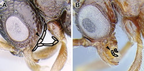
- Maxillary palps very long and reaching occipital foramen, 6-segmented and visible in mounted specimens (Figure A) (Malagasy) . . . . . Tanipone
- Maxillary palps never reaching occipital foramen, usually not visible without dissection and often with fewer than six segments (Figure B) . . . . . 11
11
return to couplet #10
- Constriction present between pre- and postsclerites of abdominal segment V, both dorsally and ventrally and helcium circumference small with helcium positioned at about the midheight of segment III (Figures A, B) . . . . . 12
- No constriction between pre- and postsclerites of abdominal segment V (Figure C) or helcium circumference large and helcium positioned above midheight of segment III. Rarely, pre- and posttergites may be separated by a gutter-like cinctus but in lateral view there is no constriction, the surface of pre- and postsclerites is contiguous and there is no cinctus on the sternites . . . . . 13
12
return to couplet #11
- Antennae with 12 segments (Indomalayan) . . . . . Eusphinctus
- Antennae with 13 segments (Neotropical) . . . . . Sphinctomyrmex
13
return to couplet #11
- Veins C and R·f3 absent from the fore wing (Figure A). Sternite of abdominal segment IX (subgenital plate) usually visible without dissection as two thin spines, in lateral view curved upwards. Posttergite of abdominal segment VIII (pygidium) often flat or impressed and delimited by a carina (Afrotropical, Indomalayan, Australasian) . . . . . Zasphinctus
- Veins C and R·f3 present in the fore wing (Figure B). Sternite of abdominal segment IX (subgenital plate) visible without dissection as two thin spines, in lateral view more or less straight or slightly upcurved. Posttergite of abdomnal segment VIII (pygidium) not delimited by a carina . . . . . 14
14
return to couplet #13
- Submarginal cell (SMC) in fore wing closed by Rs·f2–3 (a fenestra may be present at junction of Rs+M and Rs·f2–3) (Figure A) or SMC open but Rs·f2–3 present and 2rs-m also developed, closing SMC or not (Figure B) . . . . . 15
- SMC not closed by Rs·f2–3, either open (Figure C) or Rs·f2–3 completely absent and SMC closed by vein 2rs-m (Figure D) . . . . . 23
15
return to couplet #14
- Vein 2rs-m present, partial or complete in fore wing (Figure A) . . . . . 16
- Vein 2rs-m absent or at most stub-like in fore wing (Figure B) . . . . . 19
16
return to couplet #15
- Hind tibiae with one spur (Figure A) . . . . . 17
- Hind tibiae with two spurs (Figure B) . . . . . 18
17
return to couplet #16
- Marginal cell closed (Baltic amber) . . . . . Procerapachys
- Marginal cell open (Nearctic, Neotropical, Dominican amber) . . . . . Acanthostichus (in part - also #24)
18
return to couplet #16
- Mesopleuron divided by oblique groove, irregularly sculptured (Figure A) (Malagasy, Indomalayan, Baltic amber) . . . . . Chrysapace
- Mesopleuron not divided by a groove, mostly smooth with longitudinal rugae (Figure B) (Neotropical, Dominican amber) . . . . . Cylindromyrmex
19
return to couplet #15
- Costal vein (C) absent in fore wing, R·f3 absent or at most a stub past pterostigma (Figure A) . . . . . 20
- Costal vein (C) present in fore wing, R·f3 present past pterostigma (Figure B) . . . . . 21
20
return to couplet #19
- Helcium circumference large and helcium positioned supraaxially; posterior face of abdominal tergite II (petiolar node) and anterior face of abdominal tergite III poorly developed (Figure A) (Malagasy) . . . . . Lividopone (in part - also #28)
- Helcium circumference small and helcium positioned axially; posterior face of abdominal tergite II (petiolar node) and anterior face of abdominal tergite III well developed (Figure B) (Palearctic, Afrotropical, Malagasy, Indomalayan, Australasian) . . . . . Parasyscia (in part - also #26)
21
return to couplet #19
- Abdominal segment III very broadly attached to segment IV such that waist appears composed of one segment. Gaster usually widest posterior to abdominal segment IV (Figure A) (Indomalayan) . . . . . Yunodorylus (in part - also #25)
- Abdominal segment III narrowly attached to segment IV such that second segment of the waist (postpetiole) somewhat differentiated from rest of gaster. Gaster widest at abdominal segment IV (Figure B) . . . . . 22
22
return to couplet #21
- Antennal segment III the shortest segment (Figure A). In lateral view, anterior margin of eye situated very close to mandibular insertion, separated by less than maximum scape diameter. Maxillary palps with 4 segments, labial palps with 3 segments (Neotropical) . . . . . Neocerapachys (in part - also #32)
- Antennal segment II is the shortest segment (Figure B). In lateral view, anterior margin of eye is situated relatively far from mandibular insertion, separated by more than maximum scape diameter. Maxillary palp with 5 segments, labial palps with 3 segments (Indomalayan) . . . . . Cerapachys
23
return to couplet #14
- Notauli absent (Figure A) . . . . . 24
- Notauli present, at least anteriorly (Figure B) . . . . . 27
24
return to couplet #23
- Helcium circumference large and helcium positioned supraaxially; posterior face of abdominal tergite III and anterior face of abdominal tergite IV poorly developed (Figure A). Maxillary palps 2-segmented, labial palps 3-segmented (Nearctic, Neotropical, Dominican amber) . . . . . Acanthostichus (in part - also #17)
- Helcium circumference small and helcium positioned axially; posterior face of abdominal tergite III and anterior face of abdominal tergite IV developed (Figure B). Maxillary palps not 2-segmented in combination with 3-segmented labial palps . . . . . 25
25
return to couplet #24
- R·f3 vein present in fore wing (Figure A), long and conspicuous, sometimes joining Rs·f4–5 to form a closed marginal vein (Indomalayan) . . . . . Yunodorylus (in part - also #21)
- R·f3 vein absent in fore wing, at most a stub (Figure B) . . . . . 26
26
return to couplet #25
- Rs·f2–3 vein absent in fore wing. Pterostigma gives origin to a ‘free stigmal vein’ composed of 2r-rs&Rs·f4–5 (Figure A) or Rs connected to M through 2rs-m or, in smaller species, the free stigmal vein entirely absent or only a stub of 2r-rs present (Palearctic, Afrotropical, Malagasy, Indomalayan, Australasian) . . . . . Lioponera (part)
- Rs·f2–3 vein present in fore wing, long or a stub (Figure B) (Palearctic, Afrotropical, Malagasy, Indomalayan, Australasian) . . . . . Parasyscia (in part - also #20)
27
return to couplet #23
- Inner margins of antennal sockets concealed by ‘frontal carinae’ (torulo-posttorular complex) in full-face view (Figure A). Middle tibiae without spurs (Figure C) (Afrotropical, Malagasy, Indomalayan, Australasian) . . . . . Simopone
- Antennal sockets completely exposed in full-face view (Figure B). Middle tibiae with a single spur, which may be simple and inconspicuous (Figure D) . . . . . 28
28
return to couplet #27
- Helcium circumference large and helcium positioned supraaxially; posterior face of abdominal tergite II (petiolar node) and anterior face of abdominal tergite III poorly developed (similar to couplet 12 Figure A) (Malagasy) . . . . . Lividopone (part)
- Helcium circumference small and helcium positioned axially; posterior face of abdominal tergite II (petiolar node) and anterior face of abdominal tergite III well developed (similar to couplet 12 Figure B) . . . . . 29
29
return to couplet #28
- Antennae with 11 or 12 segments . . . . . 30
- Antennae with 13 segments . . . . . 31
30
return to couplet #29
- Discal cell (DC) often closed (Figure A). Abdominal segment III distinctly smaller than the succeeding segment IV, i.e. postpetiole well differentiated and often similar in size to abdominal segment II (petiole). Abdominal sternite VII almost always modified, notched, equipped with tufts of setae, with palpiform or flat projections, or otherwise hypertrophied (Figure C). Most species with 11-segmented antennae, some with 12-segmented (Indomalayan, Australasian, O. biroi is a pantropical tramp species) . . . . . Ooceraea
- Discal cell (DC) open (Figure B). Abdominal segment III may be smaller than the succeeding segment IV but usually larger than abdominal segment II (petiole). Abdominal sternite VII simple, never modified (Figure D). Antennae 12-segmented (Palearctic, Nearctic, Neotropical, Indomalayan) . . . . . Syscia
31
return to couplet #29
- Costal (C) and R·f3 veins absent from fore wing (Figure A) (Palearctic, Afrotropical, Malagasy, Indomalayan, Australasian) . . . . . Lioponera (in part - also #26)
- Costal (C) vein always present in fore wing, R·f3 often present (Figure B) . . . . . 32
32
return to couplet #31
- Rs·f2–3 vein present in fore wing (Figure A). Propodeal declivity and anterior face of abdominal segment II (petiole) surrounded by a conspicuous carina (Neotropical) . . . . . Neocerapachys (in part - also #22)
- R·f2–3 vein absent in fore wing (Figure B). Propodeal declivity and anterior face of abdominal segment II usually not surrounded by a carina (Afrotropical, Malagasy) . . . . . Eburopone


