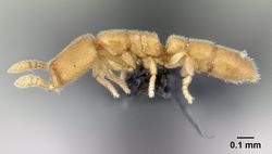
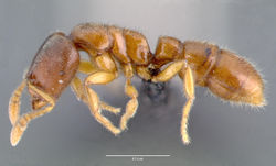
Key to Prionopelta of the Malagasy region
This worker key is based on: Overson, R. & Fisher, B.L. 2015. Taxonomic revision of the genus Prionopelta (Hymenoptera, Formicidae) in the Malagasy region. ZooKeys, 507, 115-150 (doi: 10.3897/zookeys.507.9303).
Their notes regarding the key:
The role of sculpture in identifying species of Malagasy Prionopelta
Sculpture of the head and dorsum of the mesosoma is of high diagnostic value in identifying Malagasy Prionopelta. In describing sculpture we use the same terminology as Harris (1979) and Shattuck (2008). We use the term “foveae” to refer to the shallow, circular, flat-bottomed depressions present on the integument, and the terms “punctations” or “punctures” to mean minute, point-like depressions in the surface of the integument that appear as tiny pinpricks even under high magnification. All Malagasy Prionopelta have foveae that vary in both size and density on both head and mesosoma, and most species also have punctations on the dorsum of the mesosoma. Additionally, all taxa possess a band devoid of foveae on the head that runs medially and longitudinally. This area, extending from just posterior to the antennal sockets to near the posterior margin of the head, varies in size and shape among species. Throughout the text, this area is excluded from consideration when statements are made concerning the density and extent of sculpture on the head. In some individuals a linear, scarlike, coronal suture is present in this region running longitudinally along the dorsum of the head.
When identifying Malagasy Prionopelta, it is helpful to recognize three qualitative conditions concerning the arrangement of foveae on the head. The first condition occurs when foveae are relatively equally spaced from one another at a distance that is greater than the diameter of an individual fovea, with smooth, shining integument present between foveae (Fig. 1A, B; Figures are shown in the key below). The next condition is one of variable foveal placement, with some foveae as described above but others directly adjacent to one another (Fig. 1C). At low magnification, these adjacent foveae appear to touch directly, where as under high magnification they appear very close together (much closer together than the diameter of an individual fovea), causing the area between each fovea to form a distinct raised margin that rises above what would otherwise be the smooth surface of the integument. There is a general tendency in both of the above-described conditions for cephalic foveae to increase in density moving laterally to medially in full-face view. In the final of these three general conditions, foveae are placed extremely densely on the head so that virtually no shining integument is present between foveae (except for the median cephalic band mentioned above). In this condition, foveae appear as a dense system of directly adjacent pits with only netlike boundaries of thin, raised margins separating individual fovea (Fig. 1D).
Prionopelta can be recognized from other Malagasy genera through the following combination of characters: petiole which lacks a posterior face due to its broad attachment to the gaster; elongate, subtriangular mandibles with three teeth positioned distally on a distinct mandibular face; apical tooth the longest, and second tooth the shortest with a mid-length third tooth; antennal segments variable (either 9 or 12 for Malagasy species) but always with a four-segmented club; body overall never more than 3 mm in length.
You may also be interested in
1
- Nine antennal segments present, palest and smallest of the Malagasy Prionopelta (HL < 0.4 mm and HW < 0.3 mm); entire body pale yellow . . . . . Prionopelta laurae
- Twelve antennal segments present; not as small as above (HL > 0.4 mm and HW > 0.3 mm); varying in size and color . . . . . 2
2
return to couplet #1
- Cephalic foveae widely and evenly spaced such that they are only extremely rarely adjacent to one another; all cephalic foveae appear as if scooped out of a flat, shining surface, and completely lack raised margins at their perimeter (Fig. 1A, B) . . . . . 3
- Cephalic foveae of variable spacing but never as sparse as above; at a minimum, head with several clusters of two or more foveae which are directly adjacent in full-face view (Fig. 1C) and often with many to most foveae on the head directly adjacent to one another (Fig. 1D); integument between adjacent foveae appears to bulge, and when foveae are densely placed, these bulged areas form a network of raised margins (Fig. 1D) . . . . . 4
3
return to couplet #2
- Metanotal suture absent in dorsal view, in its place a smooth surface with no clear boundary dividing mesonotum and propodeum; posterior margin of the propodeum convex and crescent-shaped in dorsal view (Fig. 2A); lamellae of the posterior propodeum present; apical tooth of the mandible extremely long (Fig. 2C) . . . . . Prionopelta vampira
- Metanotal suture present; posterior margin of the propodeum relatively straight in dorsal view (Fig. 2B); lamellae absent; apical tooth intermediate in length (Fig. 2D); known only from tropical dry forests of western Madagascar . . . . . Prionopelta xerosilva
4
return to couplet #2
- Vast majority of cephalic foveae densely positioned so they are directly adjacent to one another; vein-like ridges present running between foveae producing the appearance of a netlike pattern across the entire head (Fig. 3A, B, C, D); areas of shining integument devoid of foveae are present only at the extreme posterolateral corners of the head . . . . . 5
- Cephalic foveae variable in their spacing, but always with some foveae directly adjacent and others isolated from one another; usually with the frequency of adjacent foveae increasing laterally to medially (Fig. 1C) . . . . . 7
5
return to couplet #4
- Coronal suture present on head which appears as a uniformly thin, linear scar that swells above the surrounding integument under high magnification (Fig. 3A, B); sculpture on pronotum weak, consisting of shallow foveae which are much larger and more widely spaced than those on the head, and are interspersed with minute punctures . . . . . Prionopelta subtilis
- Coronal suture absent on head; median cephalic band wider and more irregular, never uniformly swelling above the surface of the surrounding integument as a suture, appearing instead as a shining area devoid of foveae (Fig. 3C); if band is narrow (Fig. 3D), then surface of the pronotum always covered in large, deep, regularly-spaced foveae, similar in size to those on the head . . . . . 6
6
return to couplet #5
- Cephalic foveae small and densely positioned so that the vast majority are directly adjacent to one another with swollen ridges between; median cephalic band devoid of foveae is wide, usually wider at its base and narrowing posteriorly (Fig. 3C); foveae of the pronotum much larger than that of the head, ranging from shallow to deep, and interspersed with punctures; known only from Seychelles . . . . . Prionopelta seychelles
- Cephalic foveae large, deep, and densely placed, joining to form a network of tall, jagged ridges; median cephalic band lacking foveae is usually thin and narrow, but its boundaries are irregular and jagged (Fig. 3D); pronotum lacking punctures and possessing deep, regularly-spaced foveae which are similar-sized to those on the head . . . . . Prionopelta descarpentriesi (in part - also #7)
7
return to couplet #4
- Distinctly tricolored body with dark brown, uniformly colored head, lighter brown body, and pale yellow legs; large, globular eyes which project spherically from the head; at high magnification, eyes composed of several overlapping globular sections delineated by sutures (Fig. 4A, D); known only from the Anjanaharibe-Sud Reserve of Madagascar . . . . . Prionopelta talos
- Not distinctly tricolored as above; some individuals possess dark brown on head but always at least with lighter brown present at the posterolateral corners of the head which resembles the color of the mesosoma; eyes ranging in size from almost absent (Fig. 4C, F) to dark, irregular circles that are almost flush with the surrounding integument (Fig. 4B, E); eyes never globular and never composed of several visible globular sections . . . . . Prionopelta descarpentriesi (in part - also #6)


