Myrmecina reticulata
| Myrmecina reticulata | |
|---|---|

| |
| Scientific classification | |
| Kingdom: | Animalia |
| Phylum: | Arthropoda |
| Class: | Insecta |
| Order: | Hymenoptera |
| Family: | Formicidae |
| Subfamily: | Myrmicinae |
| Tribe: | Crematogastrini |
| Genus: | Myrmecina |
| Species: | M. reticulata |
| Binomial name | |
| Myrmecina reticulata Aswaj, Anoop & Priyadarsanan, 2021
| |


The only known collection site for this species sits in the tropical semi-evergreen and evergreen forest, being a shaded spot with about 80% canopy cover in the Dampa Tiger Reserve at an elevation of 409 m above sea level. The annual rainfall ranges from 2000 mm to 2500 mm and temperature ranges from 12 °C to 25 °C during winter and 22 °C to 35 °C during summer. A single worker specimen of M. reticulata was collected using a Winkler extractor from sifted leaf litter taken from one square meter which accounted for 1000 ml of leaf litter. We were able to capture three additional ant genera: Meranoplus, Lasius and Carebara from the same trap.
Identification
Myrmecina reticulata belongs to Myrmecina gracilis complex as it has small eyes (<0.12 mm), flattened mesosoma, long and posteriorly directed propodeal spine, first tergum with concave anterior margin in dorsal view and sculptured dorsum.
The species is unique in having the following characters:
- head and mesosoma with longitudinal rugae
- anterior margin of clypeus with a prominent median tooth like process
- propodeal dorsum has a distinct short and triangular denticle located anteriorly
- propodeal spines longer than broad at base, anterodorsal margin medially elevated, slightly sloping to sides and curved up apically
- sternopostpetiolar process not well developed, blunt
- first gastral tergum reticulate
- body blackish brown
Myrmecina reticulata is close to Myrmecina bawai in the following characters:
- meosoma in dorsal view with parallel longitudinal rugae
- anterior pair of denticles present on the propodeal dorsum
- first gastral tergum in dorsal view sculptured.
However, M. reticulata can be easily separated from M. bawai by the following characteristics:
- body mainly blackish brown in M. reticulata (dorsum of head, mesosoma and gaster yellow with a dark tinge. All other body parts yellow in M. bawai)
- anterior margin of the clypeus with a distinct median tooth in M. reticulata (anterior margin of the clypeus without a median tooth in M. bawai)
- propodeal spines long, distinctly broader than long in M. reticulata (propodeal spine short, nearly as broad as long in M. bawai)
- sternopostpetiolar process not well developed, blunt in M. reticulata (sternopostpetiolar process well developed, triangular with acute anteroventral corner in M. bawai)
- first gastral tergum in dorsal view, reticulate anteriorly, coarsely reticulate posteriorly in M. reticulata (first gastral tergum anteriorly with micro-punctures, coarsely reticulate medially in M. bawai)
Distribution
Distribution based on Regional Taxon Lists
Oriental Region: India (type locality).
Distribution based on AntMaps
Distribution based on AntWeb specimens
Check data from AntWeb
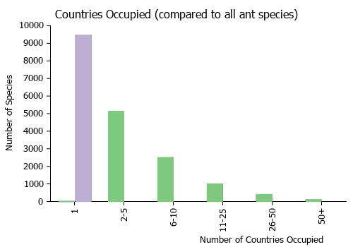
Countries Occupied
| Number of countries occupied by this species based on AntWiki Regional Taxon Lists. In general, fewer countries occupied indicates a narrower range, while more countries indicates a more widespread species. |
|
Biology
Castes
Worker
 
| |
| . | |
Nomenclature
The following information is derived from Barry Bolton's Online Catalogue of the Ants of the World.
- reticulata Myrmecina reticulata Aswaj et al., 2021: 166, figs. 4-5 (w.) INDIA.
Type Material
- Holotype: worker: “India: Mizoram, Dampa Tiger Reserve, Mamit district, (23.6948°N, 92.4283°E, 409 m), 25.iv.2019, Winkler extraction method, Coll. Punnath Aswaj and Karunakaran Anoop” (NBAIR/ HYM-FOR/9424).
Description
Worker
Head: In full face view, subrectangular with posterior margin strongly concave medially, weakly convex posterolateral margin; slightly longer than broad (Fig. 4 A). Mandibles broad, masticatory margins with large apical tooth followed by medium size preapical tooth, third tooth robust, four denticles and a distinct basal tooth (Fig. 5 A). Anterior margin of clypeus with a distinct median tooth; anterolateral corners of the median portion of the clypeus with a pair of denticles (Fig. 4 A). Antenna 12-segmented, with distinct 3-segmented club; scape not extending beyond occipital corner; antennal segment II pear-shaped; segment III–IX slightly shorter than broad; terminal segment (XII) longer than segments X and XI combined. Eyes very small, 0.05 mm in diameter, located anterolateral to mid-length of the head at a distance of 0.21 mm from the point of mandibular insertion (Fig. 4 A).
Mesosoma: In profile view, mesosoma weakly convex; anterior ventrolateral corner of pronotum dentate, but it is not developed as a spine; pronotum completely fused with mesonotum; the ventral margin of the mesopleural area is delimited from the metapleural area by a distinct notch (Fig. 4 C). Propodeum weakly demarcated anteriorly; but lack a distinct metanotal groove. In profile view, propodeal dorsum has a distinct short and triangular denticle located anteriorly; propodeal spines clearly longer than broad at base, anterodorsal margin medially elevated, slightly sloping to sides and curved up apically (Fig. 5 C). Propodeal declivity steep and shallowly concave; posterodorsal corner of propodeal lobe right angle, posteroventral margin of the lobe weakly convex (Fig. 4 C).
Metasoma: In dorsal view, petiole slightly longer than broad, almost parallel sides (Fig. 4 B). In profile view, petiole almost as long as high (excluding subpetiolar process); petiolar dorsal surface weakly concave, anterior slope nearly straight; narrow subpetiolar process with relatively pointed anteroventral corner (Figs 4 C, 5 C). In dorsal view, postpetiole broader than petiole, clearly shorter than broad, its anterior margin moderately concave while posterior margin distinctly convex (Fig. 4 B). In profile view, postpetiole almost as long as high; sternopostpetiolar process not well developed (Fig. 5 C). Gaster in dorsal view, slightly elongate circular with strongly concave anterior margin (Fig. 5 D).
Sculpture: Dorsum of head with distinct longitudinal rugae (Fig. 4 A). Ventrolateral area of head (temple + gena) with longitudinal rugae. Mandible smooth and shiny. Clypeus largely smooth (Figs 4 A, 5 A). Mesosoma with parallel longitudinal rugae, diverging towards anterior portion of pronotum (Fig. 5 B). The sculpture of the mesoand metapleural areas is distinct, with the break point aligned with the pleural suture. Dorsal surface of petiole relatively smooth anteriorly and posteriorly, punctured medially and laterally with few distinct rugae (Figs 4 B, 4 C, 5 C). Postpetiolar dorsum punctured with rugae (Fig. 4 B). All legs smooth and shiny. First gastral tergum entirely reticulate (Fig. 5 D); remaining gastral segments smooth and shiny. Pilosity: Body covered with abundant erect hairs. Antenna with abundant suberect to erect hairs. Mandibles with many suberect hairs, basal masticatory margin with few spatulate hairs. Anterolateral corner of clypeus with 4–5 relatively large erect hairs and medially with two small erect hairs. Legs with numerous suberect to erect hairs. Gaster with abundant suberect to erect hairs.
Colour: Antennae, clypeus, mandible, 2/3 of mesopleura and propodeum laterally are yellowish brown. All legs, anterolateral corner of head below eyes, gastral segments 2–4 are yellow. A distinct yellowish colour separation present laterally between blackish brown first gastral tergum and sternum. Petiole yellowish dorsally and brownish medially. Postpetiole and lateral side of petiole are yellowish brown. Cephalic dorsum, mesosoma, first gastral segments are blackish brown.
Measurements and indices (Holotype): HL—0.68, HW—0.63, MDL—0.36, EL—0.05, SL—0.47, TL—2.71 , WL—0.82, PNH—0.34, PNW—0.45, MW—0.35, PSL—0.20, PTH—0.22, PTL—0.21, PTW—0.19, PPH—0.21, PPL—0.18, PPW—0.24, CI—93, MDI—53, SI—75, PI—91, PPI—133.
Etymology
The species is named in reference to the reticulate sculpture on the first gastral tergum.